Free Lab & Molecular Biology tool
 Free Lab & Molecular Biology tool
Protein Molecular Weight Calculator
The Protein Molecular Weight Calculator determines total protein molecular mass in kilodaltons (kDa) and isoelectric point (pI) from amino acid sequence strings. Molecular weight sits behind almost every solution calculation in a protein lab - converting micrograms of enzyme into picomoles for a labelling reaction, checking whether a band on a gel matches the construct you cloned, or summing subunits for a complex. Paste a one-letter amino-acid sequence and this calculator sums the average residue masses the way Expasy ProtParam does, adds the terminal water, and corrects for disulfide bonds if you enter them. It also converts an amount in µg into nmol and molecules so you can plan stoichiometric excess directly.
Protein Molecular Weight Calculator: result
Average masses assume a typical natural-isotope composition. Intact-mass MS will read slightly different once you account for post-translational modifications (glycosylation, phosphorylation, acetylation), removal of a signal peptide, or cleavage of the initiator methionine.
At a glance
- What it does: Sums a pasted one-letter sequence into average molecular weight, with a disulfide correction and a µg-to-nmol line.
- Who it is for: Made for planning labelling excess, checking a band against a construct, and converting mass into moles.
- Typical result: The default ubiquitin sequence returns 8,564.84 Da (8.56 kDa), the value ProtParam reports for it.
How to use the protein molecular weight calculator
- Paste the mature protein sequence in one-letter codes - line breaks, numbering and a leading >FASTA header line are stripped automatically. prefill →
- Enter the number of disulfide bonds if you know it (0 for reduced/denaturing conditions). Extracellular proteins such as insulin or antibody chains lose 2.016 Da per Cys-S-S-Cys bond. prefill →
- Read the molecular weight in Da and kDa, and compare it against the 110 Da × length sanity check and your construct's expected size.
- Enter an amount in µg to convert it into nmol and molecules - the numbers you actually need for stoichiometric excess in labelling or binding reactions.
- Before trusting a precise match to MS data, check the caveats: the calculator counts exactly the sequence you pasted, nothing more.
Protein Molecular Weight: the formula & its source
A protein is a chain of amino acids joined by peptide bonds, so its mass is the sum of the residue masses plus one terminal water that the chain lost during synthesis:
MW = Σ (counti × average massi) + 18.015 Da − 2.016 Da × (disulfide bonds)
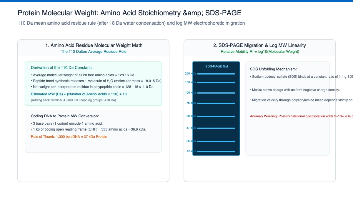
Residue masses are the average isotopic masses of each amino acid as it exists inside a chain - the free amino acid minus the water released in the peptide bond. Combining the twenty residues weighted by how common they are in proteins gives the ≈110 Da average, which is why MW ≈ 110 × length works as a quick estimate. Each disulfide bond joins two cysteine side chains with the loss of two hydrogens (2.016 Da).
| Symbol | Meaning | Unit | Typical range |
|---|---|---|---|
MW | average mass of the whole chain, water included | Da or kDa | 1,000 – 200,000 Da typical |
n | residues counted in the pasted sequence | amino acids | 10 – 1,000 |
S–S | disulfide bonds you enter by hand; each removes 2 H | count | 0 – 20 |
amount | mass to convert into nmol and molecules | µg | 0.01 – 1,000 |
Protein Molecular Weight: worked example
Worked example - human ubiquitin (the default sequence). Its 76 residues sum to 8,546.82 Da; add one water (18.02 Da) → 8,564.84 Da (8.56 kDa), matching the published ProtParam value. The 110 Da shortcut gives 76 × 110 = 8,360 Da - 2.4% low, the composition bias you would expect. For a labelling reaction, 10 µg of ubiquitin is 10 × 1,000 ÷ 8,564.84 = 1.17 nmol, about 7.03 × 10¹⁴ molecules.
Example B: disulfides on, conversion off
Bonds and a skipped conversion. Take the same 76-residue chain with disulf set to 3 and the total drops by 3 × 2.016 = 6.05 Da to 8,558.79 Da, because each bond fuses two cysteines and releases two hydrogens. A sequence with no cysteines simply ignores the field. Leaving amount at 0 skips the molar line, which suits a pure gel comparison. On a short cysteine-rich peptide the same 6.05 Da is a far larger share of the total, so bonds matter most where the chain is smallest.
How a chain of residues becomes a number
A protein is amino acids joined head to tail, and every join releases one water molecule. The calculation therefore sums the average mass of each residue as it sits inside a chain, then adds a single water back for the two free ends: MW = Σ (count × average mass) + 18.015 Da, minus 2.016 Da for every disulfide bond you enter, because each bond fuses two cysteine side chains and lets two hydrogens go.
Residue masses are average isotopic masses, weighted by how common each amino acid is. Blend the twenty in their natural proportions and a chain lands close to 110 Da per residue, which is why 110 × length works as a mental check. Paste the default ubiquitin sequence and the sum reaches 8,564.84 Da, while the shortcut gives 76 × 110 = 8,360 Da, about 2.4% low (the bias its composition predicts rather than an error in either figure).
Read the length first after pasting. A construct that comes back 2 kDa away from the size the cloning plan promised usually means the wrong sequence reached the box, and the residue count settles that argument before anything is ordered.
Mass you weigh, moles that react
Vessels are labelled in mass while reactions are written in moles, so a conversion sits between almost every protocol instruction. Enter 10 µg against ubiquitin's 8,564.84 Da and the answer is 1.17 nmol, about 7.03 × 10¹⁴ molecules; an instruction asking for a tenfold molar excess of labelling reagent now has a number to be ten times. The same division runs the other way through concentrations: 1 mg/mL of a 50 kDa protein is 1,000,000 ÷ 50,000 = 20 µM, which is the figure quoted in a binding buffer.
Stoichiometry is where mass earns its keep. One kilobase insert and a three kilobase vector at equal nanograms are a third apart in molecules, and protein work behaves the same way: a 5 µg dose of a 12 kDa enzyme carries four times the molecules of a 50 µg dose of a 60 kDa one. Convert first, then argue about ratios.
Predicted size also guards the construct. Summing subunits gives the expected mass of a complex, and a 6×His tag adds roughly 0.8 kDa, small on a gel, but visible when the same protein is compared against a mass spectrometer readout. Rough estimates can use the 110 Da rule; anything pipetted from a molar recipe wants the summed value.
What the calculated mass cannot see
The figure describes the sequence you pasted and nothing beyond it. A cleaved signal peptide, an initiator methionine trimmed by methionine aminopeptidase, N-terminal acetylation at +42 Da, phosphorylation, glycosylation running to hundreds of daltons, or a purification tag left out of the paste will all move the real mass while the arithmetic stays fixed. When intact mass differs by a few to tens of daltons, one of those explanations usually beats the calculator.
Average mass is the right number for buffers and stoichiometry. Mass spectrometry deconvolutes monoisotopic mass instead, which always reads slightly lower, so comparing the two without converting them is the classic route to declaring a clean prep wrong. Ambiguous sequence codes such as B, Z, X, J, O and U are treated as unknown rather than guessed, and disulfides are never inferred from cysteine positions. The count is exactly what you type.
Post-translational work is the boundary of the method. Insulin loses C-peptide during maturation, antibody chains carry glycans the paste cannot express, and a fusion protein must be pasted as the final construct so the junction between halves is included in the sum. The tool does the arithmetic faithfully; keeping the input honest is the reader's job.
When the number disagrees with the gel
A predicted 47 kDa protein running near 50 kDa has not necessarily failed. SDS-PAGE mobility tracks charge and shape as well as mass, glycoproteins run high, and reduced versus non-reduced lanes differ whenever disulfides keep the chain compact. Treat the calculation as the theoretical anchor and the ladder as the empirical measurement beside it.
Before rewriting a construct, recheck three inputs: the sequence actually pasted, mature form and tags handled the way reality handles them; the disulfide count suited to the conditions in the lane; and whether the amount being converted belongs to the protein you think it does. When all three agree and a gap persists, the difference is chemistry the sequence does not encode, and the next move is a measurement rather than a recalculation.
What your protein molecular weight result means
Mass is what you weigh; moles are what reacts. Every 'molar excess' instruction - a 10× excess of labelling reagent over available lysines, a 5-fold molar excess of ligase vector over insert - needs a molecular weight before it becomes a pipette volume. Worked in the other direction, MW turns a concentration into molarity: 1 mg/mL of a 50 kDa protein is 1,000,000 ÷ 50,000 = 20 µM.
It is also your fastest quality check after a construct arrives - sum the subunits to predict a complex, or predict the gel shift when a 6×His tag adds roughly 0.8 kDa.
How to read the output:| Result range | What it means | What to do next |
|---|---|---|
| Calculated mass matches the readout | sequence and construct look right | proceed with the stoichiometry |
| Off by 42 Da | an acetylation the paste missed | add the modification before comparing |
| Off by hundreds of daltons | glycosylation or a forgotten tag | check processing and the construct |
| Spectrometer reads lower | monoisotopic versus average mass | use average for solution chemistry |
| 110 × length sits nearby | composition sanity check passed | trust the summed value for pipetting |
Translate nucleotide open reading frames into predicted peptide sequences via our cDNA translation and transcription converter.
Common mistakes with the protein molecular weight
- Pasting a FASTA header, digits or line breaks along with the sequence: Headers, spaces, numbering and line breaks are stripped before counting, so check the residue length line once after pasting.
- Comparing an average mass straight against a deconvoluted spectrum: Average mass is the higher figure and monoisotopic is what the instrument reports, so convert or recalculate before judging the prep.
- Assuming the disulfide field can be inferred from cysteine positions: Bonds are never guessed from the sequence; enter the count that matches the conditions, or set zero for reduced, denatured samples.
- Leaving a retained tag out of the pasted chain: Paste the final mature sequence with any tag that stays on the protein and the sum takes care of the junction on its own.
- Reaching for a molar excess while holding only micrograms: Enter the amount to get nmol first: 10 µg of ubiquitin is 1.17 nmol, so a tenfold excess is 11.7 nmol, not 100 µg.
Limits of the protein molecular weight calculator
This is average (isotopic-average) mass, the right number for stoichiometry and buffers. Mass spectrometry deconvolutes to monoisotopic mass, which reads slightly lower.
The calculation counts exactly the sequence pasted. It cannot know about a cleaved signal peptide, an initiator methionine removed by methionine aminopeptidase, N-terminal acetylation (+42 Da), glycosylation (often hundreds of Da), or a purification tag you left out. Experimental intact mass can differ by a few Da to tens of Da for these reasons - and by much more for a glycoprotein.
Disulfide counts are entered by hand; the calculator cannot infer them from sequence. Ambiguous codes (B, Z, X, J, O, U) are treated as unknown rather than guessed.
Laboratory note from Dr. Dinesh A. Nagegowda, PhD: Predicting protein molecular weight from primary amino acid sequences (averaging 110 Daltons per residue) gives a theoretical un-modified baseline. However, in our plant biochemistry research on secondary metabolite enzymes, we observed that recombinant proteins expressed in eukaryotic systems frequently migrate larger on SDS-PAGE gels due to post-translational glycosylation, phosphorylation, or incomplete denaturation of compact hydrophobic domains.
Calculate catalytic rate constants ($k_{\text{cat}}$) from purified enzyme molecular mass using our enzyme turnover kcat calculator.
Sources
- Expasy Compute pI/MW (ProtParam), SIB Swiss Institute of Bioinformatics (used for: average amino-acid residue masses and the computed average molecular weight this calculator reproduces)
- New England Biolabs, DNA ligation with T4 DNA Ligase (protocol M0202) (used for: molar-ratio stoichiometry in macromolecular reactions, the reason mass has to become moles before a volume is chosen)
- New England Biolabs, Nucleic Acid Data (average molecular weight conventions) (used for: the average-mass-per-residue convention behind mass-to-mole conversion, the same family of arithmetic as the ≈110 Da protein average)
Cite This Calculator
For laboratory course reports, academic papers, or institutional protocols (APA format):
Protein Molecular Weight: frequently asked questions
Why does my measured mass differ from the calculated one?
The calculation is the theoretical backbone of exactly the sequence you pasted. Real proteins carry post-translational modifications, may have a processed N-terminus or removed signal peptide, and may bind buffer adducts. Compare like with like: digest your expected sequence or add the modification masses before blaming the calculator.
Should I use average or monoisotopic mass?
Use average mass for solution chemistry - molarities, stoichiometry, labelling excess. Use monoisotopic mass when interpreting intact-protein or peptide MS deconvolution, where the all-12C isotope peak is what the instrument measures. Average mass is always the higher of the two.
How accurate is the 110 Da per residue rule?
Good to a few percent for a typical protein - it is a composition-weighted average. Tryptophan-rich sequences run above it, alanine/glycine-rich sequences below. Use it as a mental sanity check, never as a substitute for the summed value in a calculation.
Does it handle tags, linkers or signal peptides?
Only what you paste. Include the tag if it stays on the mature protein; strip it if you cleave it. For a fusion protein, paste the whole final construct - the sum takes care of the junction automatically.
How do I convert µg of protein into nmol?
nmol = mass in µg × 1,000 ÷ MW in Da. The default ubiquitin example: 10 µg ÷ 8,564.84 Da × 1,000 = 1.17 nmol, about 7.03 × 10¹⁴ molecules. That is the number labelling and binding protocols mean when they ask for a 5- or 10-fold molar excess.
What does each disulfide bond subtract?
2.016 Da per bond - the two hydrogens lost when two cysteine side chains join. Reduced or denaturing conditions mean 0 bonds; native insulin or antibody chains lose 2.016 Da per Cys-S-S-Cys. The count is entered by hand because the sequence alone cannot tell you which cysteines pair.
Why is 18.015 Da added to the residue sum?
Residue masses are free amino acids minus the water each peptide bond releases, so the sum is short by one water for the intact chain. Adding 18.015 Da restores that terminal water: ubiquitin's 76 residues total 8,546.82 Da, and 8,546.82 + 18.02 = 8,564.84 Da.
What sequence formatting can I paste?
Line breaks, numbering and a leading >FASTA header are stripped automatically - paste the mature chain in one-letter codes, and only the 20 standard codes contribute. Ambiguous codes (B, Z, X, J, O, U) are treated as unknown rather than guessed, so fix or remove them before relying on the total. The calculator counts exactly what survives that cleanup.
Found a number that does not match its source? Report a mistake. Corrections are published in place and the page date is updated.
Found a number on this page that doesn’t match its source? Email the correction →.

Dr. Dinesh A. Nagegowda, PhD: Plant & Molecular Biology Editor
Dinesh is a plant molecular biologist and Chief Scientist at CSIR-CIMAP whose research runs through functional genomics, transcriptomics, metabolomics, and biosynthetic pathways in plants. He writes and…